
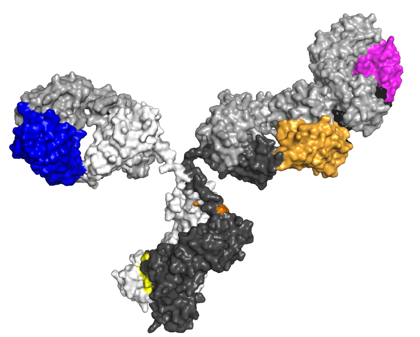
Every patient diagnosed with multiple myeloma (MM) has a unique monoclonal protein (M-protein), the abnormal protein produced by plasma cells. In the current management of MM, the M-protein is typically measured in the blood or urine using protein electrophoresis and a serum free light chain (FLC) assay. However, researchers and some medical institutions have begun to apply an age-old technology—mass spectrometry (MS)—to the diagnosis and monitoring of patients with plasma cell disorders.
“MS can be used to evaluate any complex chemical protein or subprotein in liquid samples, but the main focus of this methodology in MM is evaluating for the presence of M-protein,” explained Andrzej Jakubowiak, MD, PhD, a Professor of Medicine and Director of the Myeloma Program at the University of Chicago.
Early evidence has shown that the technique is likely more sensitive and specific than serum or urine electrophoresis or a serum FLC assay and may yield advantages for clinicians and patients. Now, researchers are working to refine the use of MS in plasma cell disorders and define and validate the ways in which it can aid in diagnosis and monitoring. Blood Cancers Today recently spoke with Dr. Jakubowiak and other experts in the field to get their insight into MS-based proteomics and its potential role in the future of MM.
Finding a New Way
For more than 50 years, gel electrophoresis has been a standard for detecting M-protein, explained Hannah Giles, MD, of the University Hospitals Birmingham NHS Foundation Trust in the United Kingdom. This technology puts a patient’s serum on an agarose gel and subjects it to an electrical current, which causes the proteins to migrate based on their electrical charge. The gel is stained with a dye and run through a light scanner. An M-protein is identified by the presence of a sharp, well-defined band with a single heavy chain. A similar band is seen with a kappa or lambda light chain.1
Serum FLC assays measure free light chains—a small part of the immunoglobulin or M-protein—and rely on evaluating the levels of the light chains and the ratio between two light chains. If there is a clonal disorder, the ratio will skew in one direction.
“Serum protein electrophoresis [SPEP] tests are not too expensive, are widely available, and have established criteria on how to monitor patients, but they have a limited degree of sensitivity,” Dr. Giles explained.
For example, in a small proportion of patients, the myeloma cells produce very small amounts of monoclonal protein. These patients are more of a diagnostic challenge, and clinicians must rely on other diagnostic tests, such as serial bone marrow tests or imaging scans, which can be painful and invasive.
Additionally, SPEP lacks specificity. A restricted gamma band can be due to causes other than M-protein, such as fibrinogen, various levels of hemolysis, or high rheumatoid factor.2 If an abnormality is detected, that sample is then sent for immunofixation (IFE), which can identify the abnormal protein.
“This method takes a while and is labor intensive,” said David L. Murray, MD, PhD, an Assistant Professor of Laboratory Medicine and Pathology in the Division of Clinical Biochemistry at the Mayo Clinic. “When I joined the lab at Mayo, we set out to try to do something new and ended up … with this MS method for detecting M-protein.”
MS Methods
MS takes samples, ionizes them into charged molecules, and measures their mass-to-charge ratio. Multiple MS methods have been developed, and researchers hope to adapt these methods for multiple uses within the diagnosis and management of plasma cell disorders.
One MS method employs immunoglobulin trypsin digestion to break proteins into peptides and detect peptides specific to the M-protein complementarity determining region (CDR); this process is sometimes called the clonotypic peptide approach, Dr. Murray said.
“We started with this method at Mayo, but it uses really expensive mass spectrometers,” Dr. Murray noted. “Ninety-five percent of these [machines] are used for research purposes and not in the clinic. I thought this was a difficult and unreliable method that required a lot of knowledge.”
Instead of using trypsin digestion, a simpler approach uses reagents and washes to isolate the immunoglobulins and remove nonimmunoglobulin proteins. The immunoglobulins are then treated to separate the light chains and spotted onto specialized plates, which are analyzed by MS. Referred to as intact light chain assays, this method can be done using liquid-chromatography mass spectrometry (LC-MS), and the assay is known as monoclonal immunoglobulin rapid accurate mass measurements, or miRAMM.2,3 Later, this method was for use with higher-throughput mass spectrometers using a form of MS called matrix-assisted laser desorption ionization time of flight (MALDI-TOF) MS.
“Instead of migrating through a gel, they migrate through a vacuum,” Dr. Murray said. “They acquire a charge and fly from the bottom of the tube to the top, and we time how long it takes to get there. That is what happens when we measure mass.”
The MALDI-TOF MS assay developed by the Mayo Clinic is often referred to as MASS-FIX in the literature, and it replaced SPEP and IFE at the Mayo Clinic in 2018. Mayo patented miRAMM and MALDI-TOF MS and licensed the methodology to Thermo Fisher under its Binding Site Division. Thermo Fisher has developed a commercialized MALDI-TOF MS assay (the brand name is EXENT, but it is also sometimes referred to in the literature as QIP-MS).
Why Better?
The MALDI-TOF MS assays are simpler and faster than traditional electrophoresis methods and appear to be more sensitive and specific.
Among the evidence in support of this approach is an early study from Mayo, which compared serum and urine MASS-FIX with SPEP, IFE, and FLC and identified 18 patients with negative results on SPEP/IFE and serum FLC who had positive M-proteins on MASS-FIX. Of the 18 patients, 10 had MM or amyloid light chain (AL) amyloidosis but were thought to have had complete hematologic response by serum/urine PEP/IFE and serum FLC.4 A larger subsequent study showed that MASS-FIX was able to identify M-protein in patients with a wide variety of plasma cell disorders.5
One validation study of the method showed that MALDI-TOF MS detected M-protein in 100% of samples that were positive by SPEP and IFE and the majority of samples that were positive by IFE but negative by SPEP. MALDI-TOF MS identified one additional positive sample among those that tested negative by both SPEP and IFE.6
“MASS-FIX is a very powerful alternative to IFE,” Dr. Jakubowiak said. “It is able to pick up an additional 20% of patients negative by IFE for presence of M-protein.”
Another advantage of MS is its ability to distinguish between a patient’s M-protein and a therapeutic monoclonal antibody.
“These days, a lot of patients will receive a monoclonal antibody, and patients with immunoglobulin G kappa monoclonal protein given a monoclonal antibody can get interference,” Dr. Giles said. “With MS you get a specific mass-to-charge ratio, allowing you to differentiate between residual disease and the therapeutic monoclonal antibody in most patients just using the standard MS assay.”
Finally, Faith E. Davies, MD, Director of the Clinical Myeloma Program at Perlmutter Cancer Center at New York University, pointed out that there is still a small percentage of patients with MM, low-grade monoclonal gammopathy of undetermined significance (MGUS), or AL amyloidosis who have a clone that cannot be detected on SPEP or serum FLC, but it can be detected on MS.
Drs. Davies, Giles, and colleagues conducted a small study in patients with nonmeasurable MM and showed that 91% of patients had detectable M-protein by MS at presentation, indicating that these patients with “nonmeasurable” disease do have low-level secretion.7 The ability to measure M-protein in these patients should allow for better monitoring of their disease.
Other Potential Uses
The International Myeloma Working Group has approved MS methodologies in lieu of IFE, and Thermo Fisher launched its EXENT Solution technology commercially in Europe in August 2023. The package includes a preparation instrument, a MALDI-TOF mass spectrometer, and software for data review.8
Additional data out of the 65th American Society of Hematology Annual Meeting & Exposition supported the use of EXENT for the diagnosis and monitoring of MM and other plasma cell disorders. One study of 460 patients showed a diagnostic sensitivity of MS of 95.0% with a diagnostic specificity of 68.2%, which the researchers said was impacted by detection of minor M-proteins not detectable on SPEP.9 MS demonstrated similar, if not superior, sensitivity compared with SPEP and a high concordance with IFE for the M-protein isotype.
In addition to its possible role of more widely replacing SPEP and IFE, MALDI-TOF is also being used to measure M-protein as a way of measuring response via measurable residual disease (MRD), Dr. Jakubowiak said.
“At the moment we usually use SPEP to monitor for response, and when we get below the detection limit for SPEP we switch to doing bone marrow tests and start using next-generation sequencing [NGS] or flow cytometry for evidence of myeloma in the bone,” said Dr. Davies. “Everyone is keen to have a blood-based marker of MRD.”
Dr. Davies said it is possible that blood-based MS techniques may be able to serve as a “bridge” between SPEP and 10−6 MRD bone marrow testing.
“We have found that certain more advanced MS techniques can be comparable [with] MRD evaluation by bone marrow-based techniques,” Dr. Jakubowiak said.
A study comparing MALDI-TOF MS in peripheral blood with next-generation flow (NGF) cytometry in bone marrow in 71 patients with MM found concordance between methods for 62% of patients (44/71); eight patients were positive using both methods and 36 were negative for both. An additional 17 patients were detectable only by MALDI-TOF, and 10 patients were detectable only by NGF.10 The samples studied were not paired samples.
Dr. Jakubowiak and colleagues also published the results of a phase II analysis where they compared MS—both LC-MS and MALDI-TOF MS—in the peripheral blood with SPEP/IFE, positron emission tomography/computed tomography imaging, multiparametric flow cytometry, and NGS in the bone marrow in 76 patients with newly diagnosed MM enrolled in a study of KRd. MALDI-TOF MS was at least as sensitive as MRD by NGS, with limit of detection of less than 10−5 in the bone marrow.11
MRD assessment using LC-MS increased sensitivity. For some paired samples LC-MS was superior to the current standard of MRD negativity, with limit of detection less than 10−5, and was at least as sensitive as MRD by NGS, with limit of detection less than 10−6.
A more recent analysis comparing MRD assessment with MALDI-TOF MS and MS-LC in peripheral blood and NGS again showed that LC-MS could possibly exceed the sensitivity of MRD by NGS in the marrow, with a sensitivity threshold of 10−6.12
“MS with or without liquid chromatography has limitations,” Dr. Jakubowiak said. “With this technique, at least at the moment, there is a requirement to have earlier samples where the M-protein is still present in reasonable abundance so you can identify the mass of M-protein. You have to have that original sample to know that the original M-protein is not detectable or substantially reduced.”
In addition, Dr. Giles said that bone marrow-based tests still appear to be the very best, but they are also more expensive and invasive for the patient, and the high negative predictive value of the MS assays is encouraging.
“There are studies looking at the sensitivity of MS compared with a bone marrow-based assay,” Dr. Giles said. “It could be that MS will become a supplement to bone marrow testing. We may wait until the patient tests negative in the blood before doing a bone marrow test.”
Remaining Questions
One question related to MS that remains is whether the method is too sensitive in certain situations.
“Anyone who presents and is suspected of MM is screened with the gel method and FLC assay, and if MGUS is picked up you have to monitor it,” Dr. Giles said. “If you are using MS and pick up something tiny, but the patient doesn’t have any suggestive symptoms, when do clinicians assign them as having this precursor condition? What are the required concentrations? Is persistence needed for a certain period of time before you label it as MGUS?”
To illustrate this point, Dr. Murray discussed a screening study of the population of Olmstead County in Minnesota that originally found that 3.2% of people aged 50 or older had MGUS.13 Subsequently, using the MALDI-TOF and miRAMM, Dr. Murray and colleagues identified a prevalence of 5.1% for MGUS among persons 50 or older.14 Most recently, using data from a clinical biobank, MASS-FIX identified MGUS in 10.8% of European American and 16.5% of Black individuals aged 50 or older.15
Whether that low-level detection of M-protein is important to each patient, and how these patients should be managed clinically, will take time to figure out.
Another pending question relates to how widely MS will be adopted.
Up until now, a main barrier to wider use has been availability, Dr. Giles said. In the United States, Mayo Clinic has MASS-FIX and would perform testing for outside institutions, but the technology is not yet commercially available.
“Now that we have the approval in Europe, people are becoming more aware of the technologies and how much they cost,” Dr. Giles said. “Moving forward, people will have to balance out how much they cost with how much information they provide.”
Another potential barrier to more widespread adoption will relate to training of laboratory staff and education of physicians who have been using SPEP for many years, according to Dr. Davies.
Ultimately, each institution is likely to weigh the learning curve and associated costs of doing MS in-house against the cost of sending samples out for testing, Dr. Jakubowiak said. “Some places, including our own high-volume lab, have felt that it would be less expensive to send out samples to the Mayo Clinic lab,” he continued. “We still get results very quickly…faster than FLC from our own lab.”
For his part, Dr. Murray is excited to see wider adoption of MS techniques for plasma cell disorders.
“I think it will be similar to what happened with microbacteria. They switched to MALDI-TOF for identifying microbacteria and that slowly progressed to be the standard,” said Dr. Murray, who admitted that he is biased in favor of the technology after seeing what it can do day in and day out.
Leah Lawrence is a freelance health writer and editor based in Delaware.
References
- Serum protein electrophoresis. Weill Cornell Medicine. Accessed December 18, 2023. https://www.myelomacenter.org/serum-protein-electrophoresis
- Murray D. Going off the gold standard: replacing gel electrophoresis with mass spectrometry. Specialty testing webinar. Mayo Clinic Laboratories. Accessed December 18, 2023. https://news.mayocliniclabs.com/2019/03/28/going-off-the-gold-standard-replacing-gel-electrophoresis-with-mass-spectrometry/
- Murray DL, Puig N, Kristinsson S, et al. Mass spectrometry for the evaluation of monoclonal proteins in multiple myeloma and related disorders: an International Myeloma Working Group Mass Spectrometry Committee Report. Blood Cancer J. 2021. doi:10.1038/s41408-021-00408-4
- Milani P, Murray DL, Barnidge DR, et al. The utility of MASS-FIX to detect and monitor monoclonal proteins in the clinic. Am J Hematol. 2017;92(8):772-779. doi:10.1002/ajh.24772
- Mellors PW, Dasari S, Kohlhagen MC, et al. MASS-FIX for the detection of monoclonal proteins and light chain N-glycosylation in routine clinical practice: a cross-sectional study of 6315 patients. Blood Cancer J. 2021;11(3):50. doi:10.1038/s41408-021-00444-0
- Mill JR, Kohlhagen MC, Dasari S, et al. Comprehensive assessment of M-proteins using nanobody enrichment coupled to MALDI-TOF mass spectrometry. Clin Chem. 2016;62(10):1334-1344. doi:10.1373/clinchem.2015.253740
- Giles HV, Cook MA, Drayson MT, et al. Redefining nonmeasurable multiple myeloma using mass spectrometry. Blood. 2022;139(6):946-950. doi:10.1182/blood.2021013794
- Thermo Fisher Scientific announces the launch of the EXENT® Solution with IVDR Certification. Binding Site. August 24, 2023. Accessed December 19, 2023. https://www.thermofisher.com/bindingsite/gb/en/news/exent-launch.html
- Berlanga O, Birtwistle J, Allen S, et al. Multi-center clinical validation of a mass spectrometry immunoassay for the diagnosis and monitoring of multiple myeloma and associated disorders. Abstract #3667. Presented at the 65th ASH Annual Meeting & Exposition; December 9-12, 2023; San Diego, California.
- Eveillard M, Rustad E, Roshal M, et al. Comparison of MALDI-TOF mass spectrometry analysis of peripheral blood and bone marrow based flow cytometry for tracking measurable residual disease in patients with multiple myeloma. Br J Haematol. 2020;189(5): 904-907. doi:10.1111/bjh.16443
- Derman BA, Stefka AT, Jiang K, et al. Measurable residual disease assessed by mass spectrometry in peripheral blood in multiple myeloma in a phase II trial of carfilzomib, lenalidomide, dexamethasone and autologous stem cell transplantation. Blood Cancer J. 2021;11(2):19. doi:10.1038/s41408-021-00418-2
- Derman B, Rosenblatt J, Avigan D, et al. P847: MRD by mass spectrometry in peripheral blood and next generation sequencing in bone marrow in a phase 2 study of daratumumab, carfilzomib, lenalidomide, and dexamethasone for multiple myeloma. Hemasphere. 2022;6:741-742. doi:10.1097/01.HS9.0000846272.91489.2d
- Kyle RA, Therneau TM, Rajkumar V, et al. Prevalence of monoclonal gammopathy of undetermined significance.
N Engl J Med. 2006;354:1362-1369. doi:10.1056/NEJMoa054494 - Murray D, Kumar SK, Kyle RA, et al. Detection and prevalence of monoclonal gammopathy of undetermined significance: a study utilizing mass spectrometry-based monoclonal immunoglobulin rapid accurate mass measurement. Blood Cancer J. 2019. doi:10.1038/s41408-019-0263-z
- Vachon CM, Murray J, Allmer C, et al. Prevalence of heavy chain MGUS by race and family history risk groups using a high-sensitivity screening method. Blood Adv. 2022;6(12):3746-3750. doi:10.1182/bloodadvances.2021006201


 © 2025 Mashup Media, LLC, a Formedics Property. All Rights Reserved.